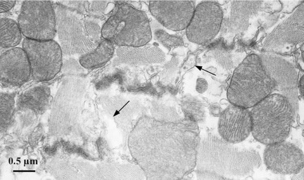
Figure 6.

Electron micrograph (TEM) section of a LFN-exposed ventricular myocardium, showing a typical steplike intercalated disc and numerous mitochondria surrounding the sarcomeres. Cell membrane separation (arrows) is observed in the interplicate region of the intercalated disc.
