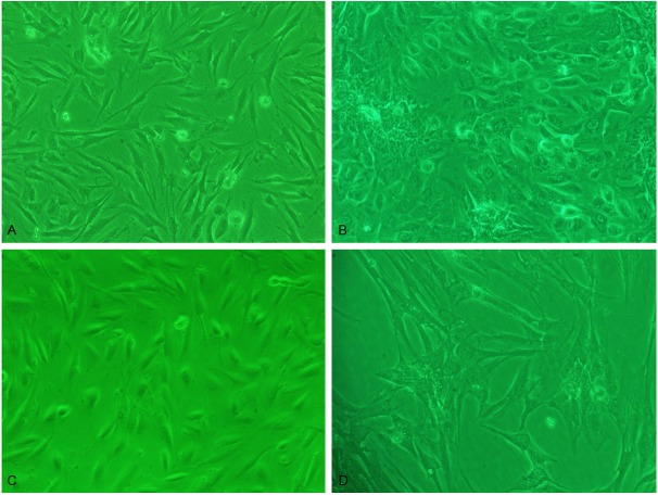
Figure 1.
ESC morphology was observed under inverted microscope: A. Eutopic ESC of primary culture 6 d. The cell was more like the fibroblasts cell (×100); B. Ectopic ESC of primary culture 6 d. The cell was mainly polygonal cell (×100); C. Eutopic ESC of the passage 3 culture 6 d. Cell was spindle and in vortex state (×100); D. Ectopic ESC of the passage 3 culture 6 d. Cell was large and in flat state (×100).

