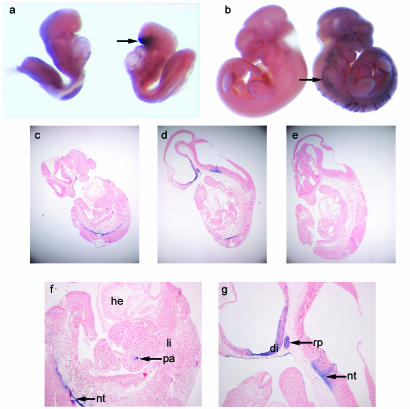
FIG. 1.
Cre recombinase expression in Z/AP+/Rip-cre+ embryos. (a and b) AP whole-mount staining of Z/AP+/Rip-cre+ embryos (right) and Z/AP+/Rip-cre− littermates (left). (a) Embryo at 8.5 dpc showing Cre expression (purple AP staining) in the region of Rathke's pouch (arrow). (b) Embryo at 11.5 dpc showing scattered staining of cells in the lower dorsal region of the embryo and the vagal (X) and accessory (XI) cranial nerve trunk (arrow). (c to g) Sections from 11.5-dpc embryos stained with AP as whole mounts (as above). Sections were counterstained with NFR. (c and d) Z/AP+/Rip-cre+ embryos showing AP staining (magnification, ×40). (e) Z/AP+/Rip-cre− littermate control. (f and g) Z/AP+/Rip-cre+ embryos (magnification, ×100) showing AP staining in the pituitary primordium, i.e., Rathke's pouch (rp), the diencephalon (di), the floor plate of the neural tube (nt), and scattered cells which may correspond to pancreatic precursor cells (pa). Liver (li), heart (he), and other tissues are not stained by AP.

