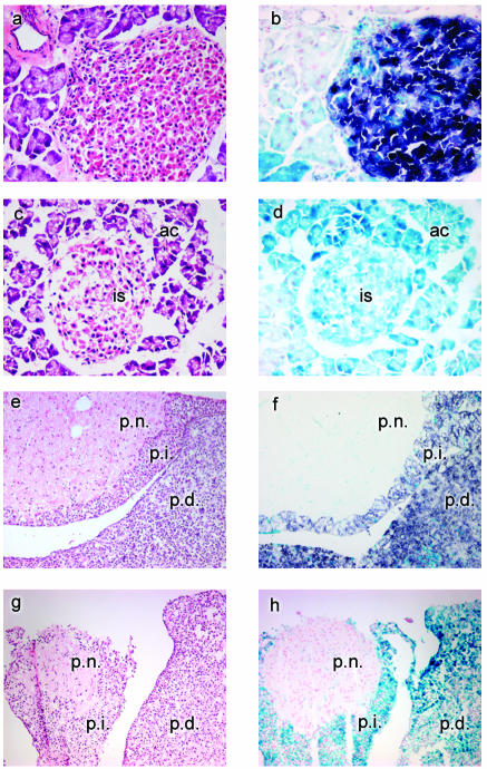
FIG. 2.
Detection of Cre-mediated excision. Pancreatic and pituitary tissues (magnification, ×400) from Z/AP+/Rip-cre+ (a, b, e, and f) and Z/AP+/Rip-cre− (c, d, g, and h) mice were examined. Tissues stained with X-Gal and AP are indicated by blue staining and purple staining, respectively. Hematoxylin and eosin staining of parallel sections for all tissues is shown in panels a, c, e, and g. (b) Islet cells showing mosaic expression of the lacZ reporter gene. Cre excision is indicated by AP staining. X-Gal staining is present in surrounding acinar cells. (d) Pancreatic section showing expression of the lacZ reporter transgene only. is, islet cells; ac, acinar cells. (f) Pituitary section showing extensive Cre activity (AP staining) in the pars distalis (p.d.) and pars intermedia (p.i.). Note that only lacZ expression is faintly detected in the pars nervosa (p.n.). (h) Pituitary section counterstained with NFR and showing expression of the lacZ reporter transgene only.

