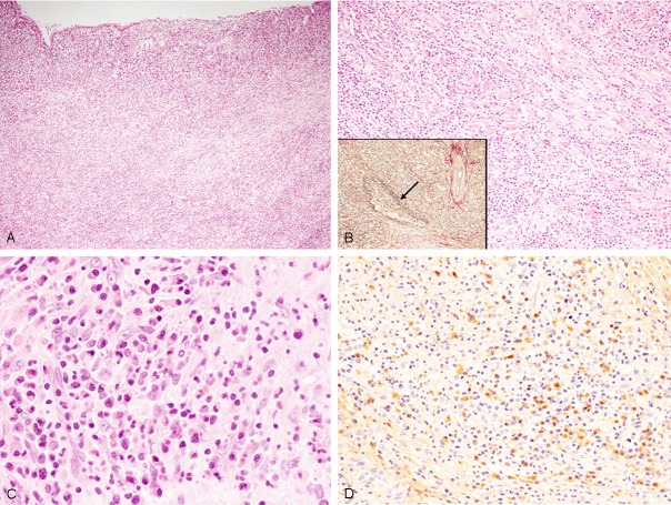
Figure 3.
Histopathological and immunohistochemical features of the gallbladder. A: Dense lymphoplasmacytic infiltration is observed in the entire gallbladder wall, HE, x 40. B: Dense lymphoplasmacytic infiltration with fibrosclerotic change is noted, HE, x 100. Obliterative phlebitis (arrow), elastica van Gieson staining x 100. C: Lymphocytes and plasma cells appear mature and are without atypia. Eosinophils are also observed, HE, x 400. D: Many IgG4-positive plasma cells have infiltrated into the lesion, x 200.

