FIG. 1.
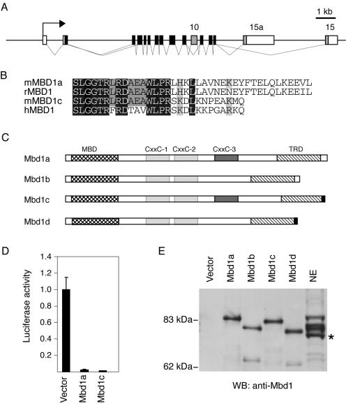
Mbd1 isoforms in mouse. (A) Schematic of the murine Mbd1 locus showing the differentially spliced exons (exons 10, 15, and 15a) shaded in gray. (B) Alignment of the C termini of the predicted MBD1 proteins from the human MBD1v1 (hMBD1), rat MBD1 (rMBD1), and murine Mbd1a/b (mMBD1a) and Mbd1c/d (mMBD1c) cDNA. (C) Schematic representation of the predicted Mbd1 proteins in mouse. CXXC domains 1 and 2 are shown shaded in light grey; the differentially spliced CXXC-3 domain is shaded in dark gray. The MBD (cross-hatched) and TRD (hatched) are indicated, and the novel alternative C terminus is shaded in black. (D) Mbd1a and Mbd1c isoforms both repress a methylated reporter gene. HeLa cells were transfected with the methylated pGL2 reporter and expression vectors for Mbd1a or Mbd1c. Means and standard deviations of the luciferase activity of triplicate transfections from a representative experiment are shown. The value for the activity without an Mbd1 expression vector is arbitrarily set to 1. (E) A Western blot (WB) of in vitro transcription and translation reactions of the possible splice variants alongside a sample of nuclear extract (NE) from murine fibroblasts. Bands in the NE lane are somewhat bowed due to the high protein concentration and are therefore aligned at their leading edges. The asterisk marks a cross-reacting band that is not Mbd1.

