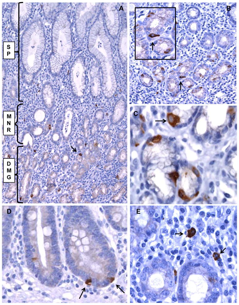
Figure 1. Lgr5 expression in gastric mucosa detected by immunohistochemistry.
A. H. pylori positive gastric antral mucosa with multiple Lgr5-positive cells in the gastric glands (arrow). The gastric pit and surface epithelium (SP), gland mucous neck region (MNR), and deep mucous gland region (DMG) are indicated. B. Lgr5-positive epithelial cells in gastric antrum from another case (arrow). C. Lgr5-positive cells showing triangular shape and basal orientation (arrow) in the glandular profiles. D. Lgr5-positive cells (arrows) in a focus of intestinal metaplasia. E. Rare Lgr-5 positive cells in the lamina propria (arrows). A and B (original magnification 200X), C, D, E (original magnification 400X).

