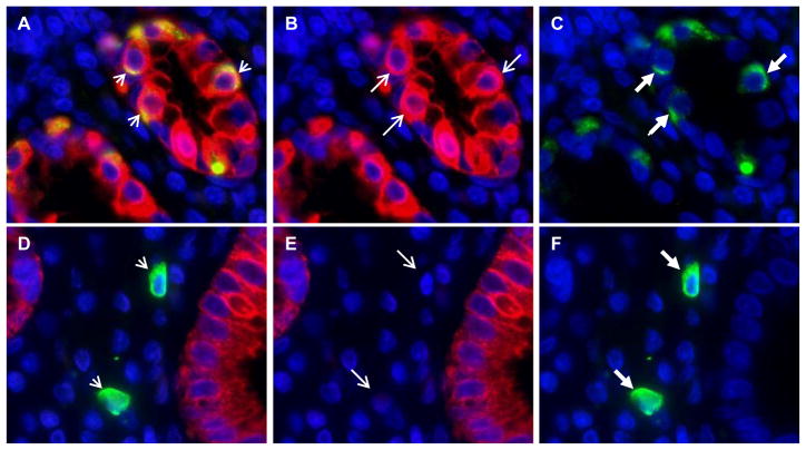
Figure 3. Co-localization of Lgr5 and cytokeratin in epithelial cells of gastric antral glands.
Panel a: Overlapped immunofluorescence images show co-localization of Lgr5 and cytokeratin in epithelial cells (yellow cells, short arrow). Panel a was created by overlapping the cytokeratin AE1/AE3 immunofluorescence stain (shown in panel b), where red stain and the long thin arrow indicate cytokeratin positive cells, with the Lgr5 immunofluorescence stain (shown in panel c), where green stain and the long thick arrow indicate Lgr5-positive epithelial cells. Panel d: Overlapped immunofluorescence images show lack of co-localization of cytokeratin in Lgr5-positive lamina propria cells (short arrows). The overlapped images in panel d were the cytokeratin immunofluorescence stain (shown in panel e), and the Lgr5 immunofluorescence stain (shown in panel f). Two Lgr5-positive lamina propria cells are indicated by the long thick arrows in panel f, and these two cells are negative for cytokeratin (panel e, long thin arrows). Nuclei are stained with DAPI. Original magnification 400X.

