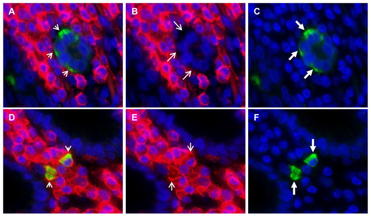
Figure 4. Lgr5-positive cells in lamina propria of gastric antrum co-express CD45.
Panel a. Overlapped immunofluorescence images show lack of co-localization of CD45 in Lgr5-positive epithelial cells (short arrows). Panel a was created by overlapping the CD45 stain (shown in panel b) with the Lgr5 stain (shown in panel c). Lgr5-positive epithelial cells stain green and are indicated by long thick arrows in panel c. The CD45 stain is negative in Lgr5-positive epithelial cells, as indicated by the long thin arrows in panel b. Panel d was created by overlapping the CD45 stain (shown in panel e) with the Lgr5 stain (shown in panel f) and shows co-localization of CD45 in Lgr5-positive lamina propria cells (yellow cells, short arrows). Lgr5-positive lamina propria cells stain green and are indicated by long thick arrows in panel f. The CD45 stain is positive in Lgr5-positive lamina propria cells, as indicated by the long thin arrows in panel e. Nuclei are stained with DAPI. Original magnification 400X.

