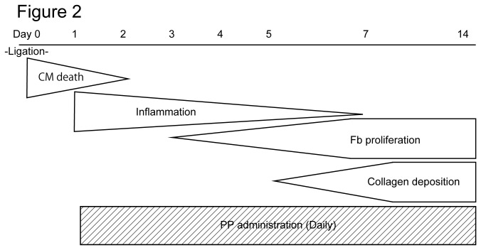
Figure 2. Schematic diagram of the pathological processes of the infarcted heart and the PP administration protocol.
Within 24 hr following the ligation (or occlusion) of left coronary artery, most of cardiomyocytes (CM) die, which is followed by inflammation and fibrosis. Fibrosis is caused by proliferation and migration of cardiac fibroblasts (Fb) beginning at approximately day 3 post ligation (occlusion). These cardiac fibroblasts secrete collagen that forms collagen fibers and become deposited in the tissue and forms scar. The collagen deposition peaks at day 7 - 14 post-ligation (occlusion). PP was administered daily to mice after 1 day following the ligation of left coronary artery (i.e. after most of cardiomyocytes die).

