
Figure 3. Decreased cell proliferation in the PP-treated infarcted heart in a mouse model of myocardial infarction.
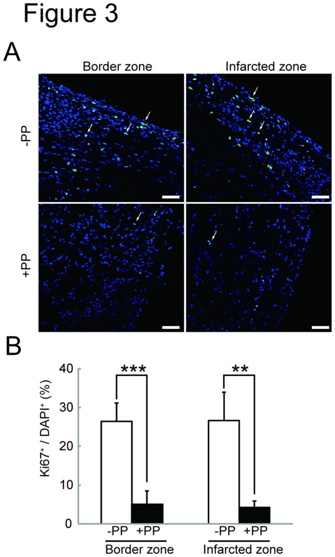
A. An effect of PP on cell proliferation in the infarcted heart. Ki67+ (green) and DAPI+ (dark blue) cells are identified by immunofluorescence. The proliferating Ki67+DAPI+ (light blue) cells in both border and infarcted zones of vehicle (DMSO) (-PP) or PP (+PP) treated mice are indicated (arrows). Scale bars: 50 μm. B. Quantitation of Ki67+ proliferating cells. The number of Ki67+ proliferating cells was counted from sections from mice (-PP: n=3; +PP: n=4) counted and shown as % (Ki67+ / DAPI+).