
Figure 5. Reduced fibrosis in the PP-treated infarcted heart in a mouse model of myocardial infarction.
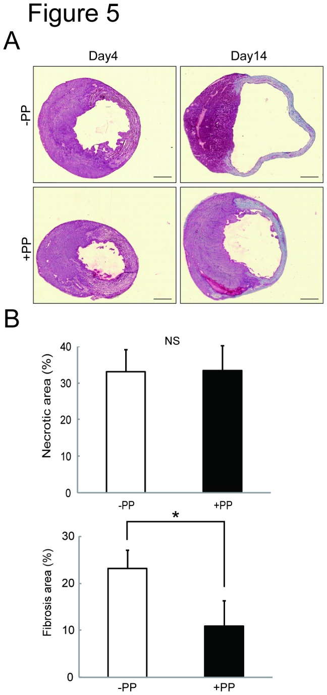
A. Masson’s trichorme stained heart sections at day 4 and day 14 post-ligation. Scale bars: 1 mm. B. Quantitation of infarct size and fibrosis area. The infarct size was measured by quantitating the necrotic area of the heart sections prepared from the vehicle treated (-PP) (n=3 mice) and PP treated (+PP) (n=4 mice) mice at day 4 post-ligation, and shown as % (necrotic area / whole heart area) (left). The fibrosis size was measured by Masson’s trichrome staining method using the heart sections prepared from the vehicle treated (-PP) (n=3 mice) and PP treated (+PP) (n=4 mice) mice at day 14 post-ligation, and shown as % (fibrosis area stained blue / whole heart area).