
Figure 2.
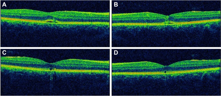
Optical coherence tomography. (A and B) Disruption of the outer retinal layers with nonspecific retinal thickening in both eyes at presentation. (C and D) Disruption of the inner segment/outer segment junction in both eyes, at 3 months after presentation.
Note: A and C are right eye; B and D are left eye.