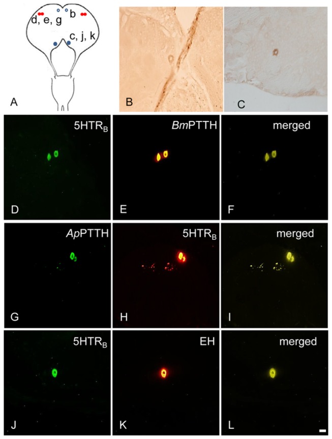
Figure 5. Colocalization of 5HTRB- and PTTH-ir in the erly pupal BR-SOG of.

A. pernyi. BmPTTH-ir/ApPTTH-ir in the 5-day-old pupal brain of A. pernyi and its colocalization with Ap5HTRB-ir (red filled circles). BmEH-ir in the 5-day-old pupa brain and its colocalization with Ap5HTRB-ir (blue filled circles) and unique distribution (open circles) of BmEH-ir in other regions of the brain. (A) The loaction of detected cells. Lower-case letters correspond to the regions shown in the photographs (e.g., b to B). (B) One BmEH-ir neuron in the PI. (C) BmEH-ir in the DC region. (D, H) 5HTRB-ir in the DL region. (E) BmPTTH-ir in the DL region. (F) Merged image of BmPTTH-ir and 5HTRB-ir in the DL region. (G) ApPTTH-ir in the DL region. (I) Merged image of ApPTTH-ir and 5HTRB-ir in the DL region. (J) 5HTRB-ir in the DC region. (K) One EH-ir neuron in the DC. (L) Merged image of BmEH-ir and 5HTRB-ir in the DC region. Scale bar = 100 µm.
