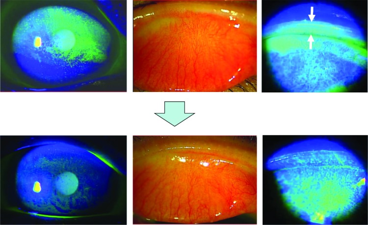
Figure 1.
A 77-year-old female. Slit lamp microscopy with fluorescein staining showed diffuse corneal erosion in the superior cornea (upper left) and lid wiper staining (upper right, white arrows) with hyperemia of palpebral conjunctiva in the right eye (upper middle). Fluorescein staining of the cornea and lid margin was improved in 2 weeks after administration of topical rebamipide eye drops (lower images).

