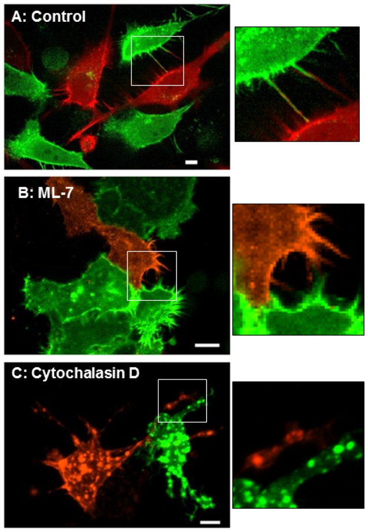
Figure 6. Micrographs of cells grown in collagen gels.
HeLa cells were transfected with Lifeact mCherry (red) or Lifeact-GFP and grown in collagen gels. These gels were not released from the walls of the wells to prevent motion artifacts. Panel A shows control (untreated) cells extending filapodia that contact neighboring cells (also see movie in Figure S2). Panels B & C show cells that were treated with 20 μM ML-7 (B) or 6 μM cytochalasin D (C). Cells treated with ML-7 continue to actively extend filopodia. Cells treated with cytochalasin D stop extending filopodia and the actin in these cells appears to collect in large aggregates. The insets are blow ups of the boxed areas. Size bar = 10 μm.

