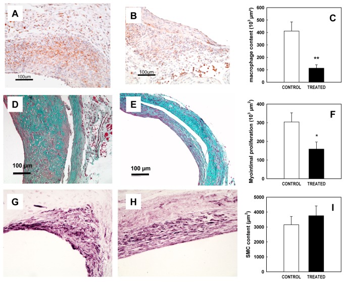
Figure 1. Effect of SSR128129E on neointimal proliferation in the vein graft model.
A-C: Effect on lesion macrophage content 2 weeks after surgery
Representative sections of vein grafts immunostained for macrophages (mac3) in control (A) and SSR128129E-treated mice (B). The size of the regions showing macrophage infiltration was determined by image analysis (C, n= 4 per group).
D-I: Effect on neointimal proliferation 8 weeks after surgery.
Representative sections of vein grafts stained by Masson’s trichrome in control (D) and SSR128129E-treated mice (E). The area of neointimal proliferation was determined by image analysis (F, n= 8-11 per group). Smooth muscle content is shown in representative sections of vein grafts labelled for α-actin in control (G) and SSR128129E-treated mice (H). Smooth muscle content as determined by the area of α-actin staining was determined by image analysis (I, n= 8-11 per group).

