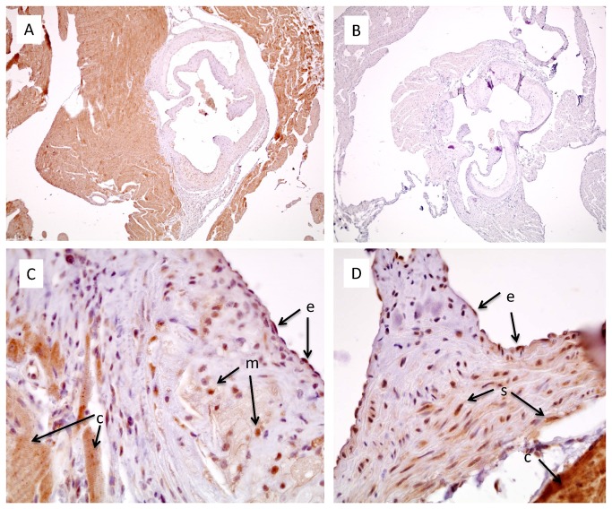
Figure 4. FGFR1 Immunostaining in the aortic sinus of 6 month-old mice.
Sections from apoE-deficient (A,B,C) or C57BL/6 mice (D) were immunostained for FGFR1. Omission of the primary antibody led to complete disappearance of the staining (B). The labels next to the arrows indicate the different cell types: e - endothelial cells; m – macrophages; c – cardiomyocytes; s – smooth muscle cells. A 4x objective was used for wide field images (A,B), whereas a 40x objective was used for the high resolution photomicrographs (C,D).

