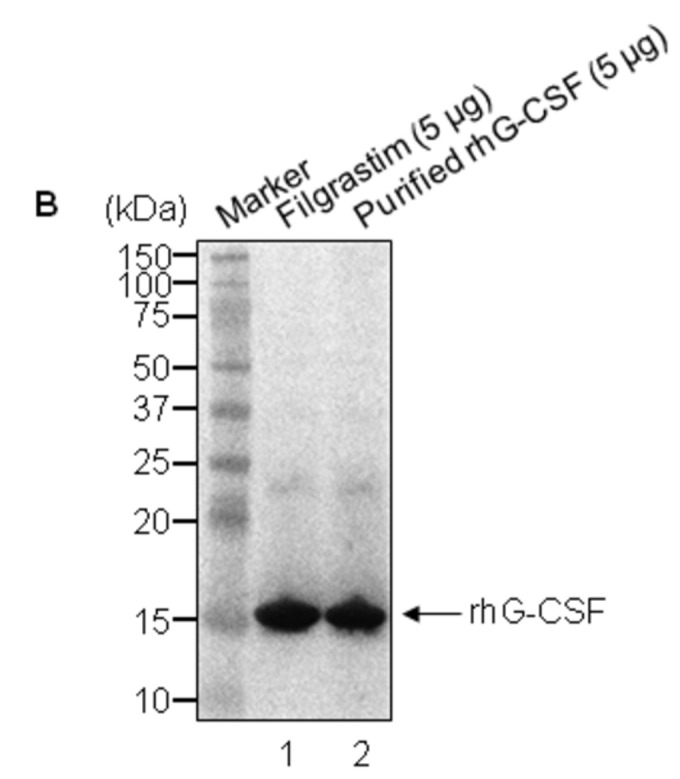
Figure 7. Peptide map and Western blot analysis of standard hG-CSF and purified rhG-CSF.

A. Two chromatograms of standard hG-CSF and rhG-CSF are overlapped for comparison. The solid arrow is the chromatogram for rhG-CSF and the dotted arrow is the chromatogram for standard hG-CSF. Absorbance is in absorbance units (AU). B. Two rhG-CSF proteins were examined by western blot after a 4–12% reducing SDS-PAGE performance. Lane 1, standard rhG-CSF (Filgrastim, 5 μg); lane 2, purified rhG-CSF (5 μg). The arrow indicates rhG-CSF.
