
Figure 7. Co-culture with HO-1-induced PC3 cells increased HO-1 expression in bone explants.
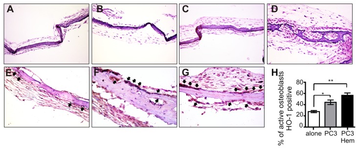
Upper panel. H&E staining of representative neonatal mouse calvariae cultured in vitro in the absence (A) or presence of PC3 cells control (B) or PC3 cells pre-treated with hemin (C). Insulin was used as a positive control of bone formation (D). Lower panel. Immunohistochemical staining of HO-1 of representative neonatal mouse calvariae cultured in vitro in the absence (E) or presence of PC3 cells control (F) or PC3 cells pre-treated with hemin (G). The arrows indicate the positive immunostaining in active osteoblasts. Magnification, x 250. The bands indicate the percentage of HO-1 positive osteoblasts respect to total active osteoblasts (H) (Significant difference, * P<0.05; ** P<0.01).