
Figure 8. Histological analysis of intra-osseous metastasis.
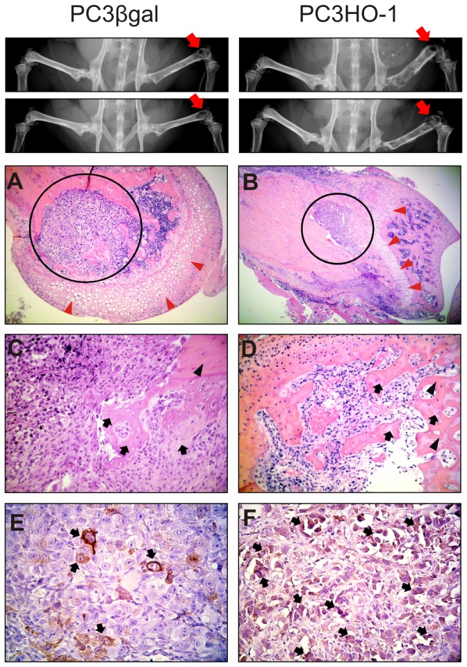
PC3HO-1 cells and PC3βgal were injected into distal head of the right femur of SCID mice. Upper panel, X-ray imaging of mouse legs after intrabone injection of PC3HO-1 or PC3βgal cells. Red arrow, injected limb. Lower panel, longitudinal section of mouse femur stained with H&E (A&B, magnification x 100; C, magnification x 400; D, magnification x 250). The red arrow heads indicate cartilage; black circle, tumor; black arrows, immature bone and black head arrows mature bone. Immunohistochemistry of HO-1 (E&F, magnification x 400) with scattered cells positive in cytoplasm (black arrows).