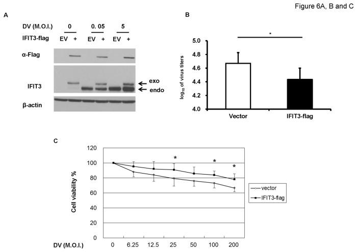
Figure 6. Overexpression of IFIT3 significantly enhanced cell survival and blocked DV replication.
A549 transfected with IFIT3-flag or empty vector (EV) for 24 h were infected by mock or DV at M.O.I. = 0.05 or 5 for 48 h. The expression of endogenous and exogenous IFIT3 was determined by western blotting as described in the Materials and Methods (A). The supernatants were collected for determining virus titers by plaque assays (B). After transfection, the cell were reseeded onto 96 well plate overnight and then infected by mock or DV at various M.O.I. = 6.25 to 200 for 48 h, the cell viability was determined by MTT assay (C). The representative results and the analysis pooled from at least three independent experiments are shown. The analysis was performed by student’s T test (B) or ANOVA (C) as described in Materials and Methods. *P<0.05.

