
Figure 4. hAIM increases the intracellular killing of M. tuberculosis without modifying the phagocytic capacity of MФ.
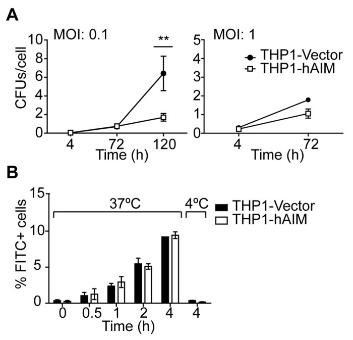
A) Stably transfected THP1 MФ were infected with M. tuberculosis at MOI 0.1 and 1. Four, 72 and 120 h later, cells were lysed and intracellular CFU numbers were determined by serial dilutions on 7h9 agar plates. CFUs per cell were calculated by dividing CFUs by number of viable cells determined by crystal violet staining at each time point. Mean ± SEM from three independent experiments performed in duplicate. **p≤0.01; *p≤0.05 two-way ANOVA. B) THP1 MФ were incubated with FITC-labelled bacilli at MOI 40 and the percentage of FITC-positive cells at the indicated time points and temperature was determined by flow cytometry. Results are expressed as the % of FITC-positive cells at each time point and show the mean ± SEM from three independent experiments.