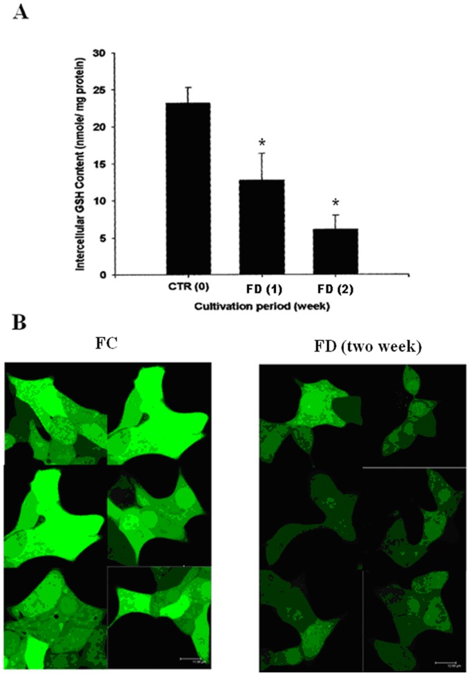
Figure 3. Folate deficiency triggers intracellular GSH depletion in RINm5F cells.
(A) The fluorometric o-pathalaldehyde (OPT) method was used to quantify intracellular GSH contents in RINm5F cells cultivated under either FC or FD condition. At the end of cultivating cells under FD condition for 2-week period, total GSH contents were found to be depleted 75% as compared to their FC counterparts. Data shown are mean ± standard deviation of triplicate determinations. *p<0.05 vs. control. (B) Probe-based confocal microscopic imaging technique was used to confirm GSH depletion phenomenon instigated by FD. Under similar cultivation period (2-week), a drastic loss of the green fluorescence of cellular CMF-GSH conjugate associated with folate-deprived cells was a direct testimony of intracelluar GSH depletion.

