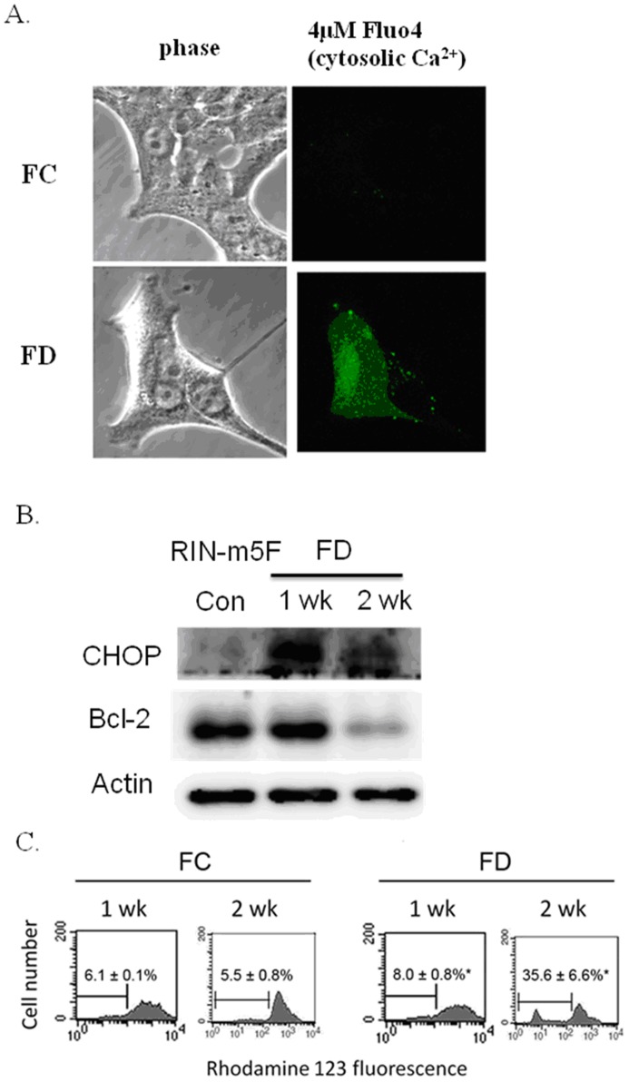
Figure 4. Folate deficiency induces cytosolic calcium (Ca2+) overload and causes disruption of △Ψm in RINm5F cells.
(A) Probe-based confocal microscopic imaging technique was used to detect cytosolic Ca2+ overload. Under FC condition, cytosolic Ca2+, as reflected by the green fluorescence of Fluo-4 probe, was essentially non-detectable. Conversely, as a result of cultivating cells under FD condition for 2-week period, a cytosolic Ca2+ overload phenomenon could be detected. This phenomenon may be attributed to the depletion of ER Ca2+ by NF-κB-dependent iNOS-mediated overproduction of NO. (B) The interplay of NO and cytosolic Ca2+ provoked ER stress as evident by the activation of CHOP expression in RINm5F cells cultivated under FD condition. (C) △Ψm was evaluated by using the rhodamine 123 fluorescent dye and meansured flowcytometrically. The disruption of △Ψm, as reflected by the percentage of cells with depolarized mitochondrial membrane, was much higher in FD cells (35.6±6.6%) when compared to FC cells (5.5±0.8%). Apparently, FD could sensitize cells to undergo apoptosis. Values shown are mean ± standard errors of 3 independent experiments. *p<0.05 vs. control.

