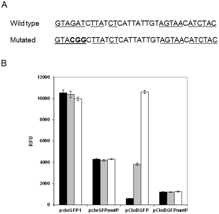
Figure 2. Mutagenesis analysis of the putative protein binding site within chr promoter region.
A) Imperfect inverted repeat of the original and mutated sequence. The mutated nucleotides are shown in bold. B) Fluorescence of E. coli carrying the mutated and no-mutated plasmid constructs. Fluorescence was measured after 3 h of growth in medium without chromate (black bars), with 1 µM (grey bars) and 10 µM (white bars) of chromate. The values represent averages and standard deviations of three replicates.

