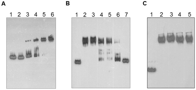
Figure 6. Gel mobility shift assays.

A) Band shift assays were performed by incubating the DNA fragment (chr promoter) with increasing concentration of ChrB protein. Lane 1, competitor protein (EBNA extract from Pierce); lane 2, 0 µM ChrB; lane 3, 1 µM ChrB; lane 4, 3 µM ChrB; lane 5, 10 µM ChrB; lane 6, 30 µM ChrB. B) DNA fragment was incubated with ChrB (10 µM) in the absence and in the presence of unlabeled competitor DNA. Lane 1, without protein; lane 2; 0 µg/µl; lane 3, 1 µg/µl; lane 4, 10 µg/µl; lane 5, 50 µg/µl; lane 6, 100 µg/µl; lane 7, 250 µg/µl of competitor DNA. C) EMSA assays with or without chromate. The chr promoter was incubated with ChrB protein (10 µM) and with increasing concentrations of chromate. Lane 1, without protein (control); lane 2, without Cr(VI); lane 3, with 10 µM Cr(VI); lane 4, with 100 µM Cr(VI); lane 5, with 1 mM Cr(VI).
