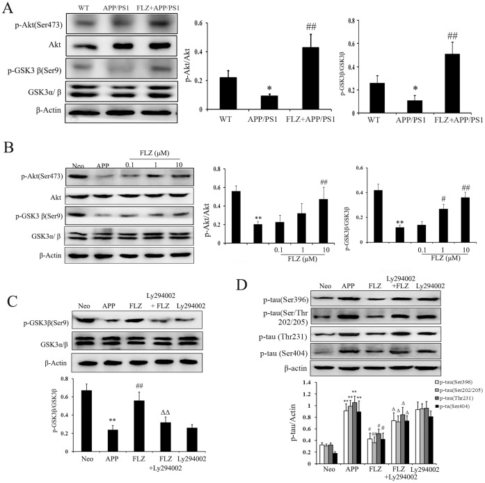
Figure 8. FLZ attenuated tau phosphorylation through regulating Akt/GSK3β pathway.
APP/PS1 double transgenic mice were orally treated with FLZ 150 mg/kg for 20 weeks. SH-SY5Y (APPwt/swe) cells were grown in “stimulating medium” containing 50% DMEM, 50% Opti-MEM, 0.5% FBS, 200 µg/ml G418 and 10 mM butyric acid sodium salt for 12 h to induce the transgene expression. FLZ (0.1, 1 and 10 µM), Ly294002 10 µM combined with FLZ 10 µM or alone were incubated with cells for 24 h. (A) Western blot assay of p-Akt (Ser473), Akt, GSK3β and p-GSK3β (Ser9) in hippocampus of APP/PS1 mice. A representative immunoblot from 4 mice was shown. Results were expressed as mean ± SD. *P<0.05 vs. WT mice; # P<0.05, ## P<0.01 vs. APP/PS1 mice. (B) Western blot assay of p-Akt (Ser473), Akt, GSK3β and p-GSK3β (Ser9) in SH-SY5Y (APPwt/swe) cells. A representative immunoblot from four independent experiments was shown. Results were expressed as mean±SD. **P<0.01 vs. Neo SH-SY5Y cells, # P<0.05, ## P<0.01 vs. solvent-treated SH-SY5Y (APPwt/swe) cells. (C,D) Ly294002 was added to test the effect of Akt on GSK3β activity and tau phosphorylation in SH-SY5Y (APPwt/swe) cells treated with FLZ. A representative result of three independent experiments was shown. Results were expressed as mean±SD. **P<0.01 vs. Neo SH-SY5Y cells, # P<0.05, ## P<0.01 vs. solvent-treated SH-SY5Y (APPwt/swe) cells; Δ P<0.05, ΔΔ P<0.01 vs. FLZ-treated SH-SY5Y (APPwt/swe) cells.

