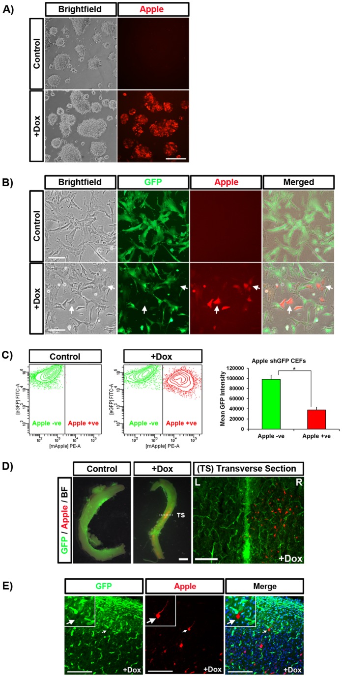
Figure 2. Induced transposon expression in mouse ES cells, CEFs, and embryos.
(A) Mouse ES cells stably transfected with PB Tet-On Apple shGFP and treated with dox for 72 hours. (B) GFP+ CEFs stably transduced with PB Tet-On Apple shGFP and treated with dox for seven days. In Apple+ cells, GFP expression was visually reduced. (C) Flow cytometry confirmed that GFP fluorescence was significantly lower in Apple+ cells, * p<0.05. (D) PB Tet-On Apple shGFP was electroporated into the neural tube of stage 14HH GFP+ embryos. Seven days post-electroporation, the embryos were treated with dox for seven days. After dissection, Apple protein is visible in the electroporated section of the spinal cord in embryos treated with dox. (E) Confocal microscopy of a transverse section revealed Apple+ neurons with reduced GFP fluorescence. L = left, R = right. Scale bars: A, B = 50 µm, D middle = 1 mm, D right = 100 µm, E = 200 µm.

