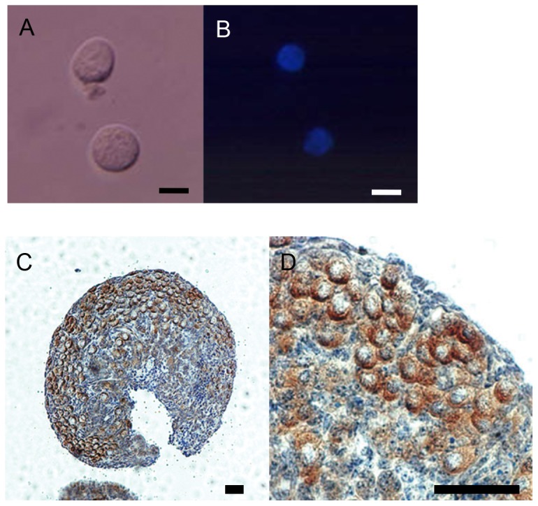
Figure 1. Microphotographs of non-growing oocytes.

(A, B) Bright and fluorescent with Hoechst 33342 stain fields. The oocytes were isolated from the ovaries of newborn pups at day 0 of delivery. Bar=10 µm. (C, D; a higher magnification of C) The ovary subjected to immunostatining for MVH. The ovary is occupied by primary oocytes that are MVH positive in the oocyte nests. Bar=50 µm.
