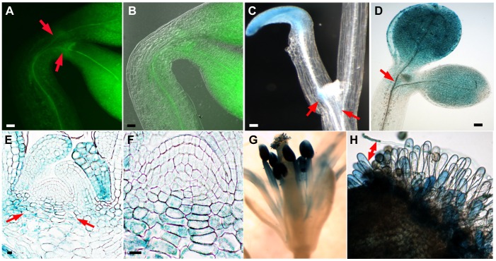
Figure 1. GUS and GFP reporter gene analyses of NDL1 expression around the vegetative and reproductive SAMs in Arabidopsis seedlings.
(A) NDL1-GFP localization in three-day-old etiolated seedlings. GFP fluorescence is detectable in the vicinity of the SAM. (B) Panel A with an overlay of the DIC image. (C) and (D) GUS histochemical staining in eight-day-old etiolated seedlings (C) and in ten-day-old seedlings grown in the light (D). GUS staining is not detectable in the SAM. (E) GUS histochemical staining in a longitudinal section of the vegetative SAM in etiolated seedlings. (F) Enlarged view of the SAM from panel E. (G) GUS staining in a mature stamen. (H) Papillar cells showing GUS staining upon pollen germination (the red double-ended arrow points to a germinating pollen tube). Scale bars = 50 µm, Red arrows in panels A, C, D and E indicate cell zones at the periphery of the SAM.

