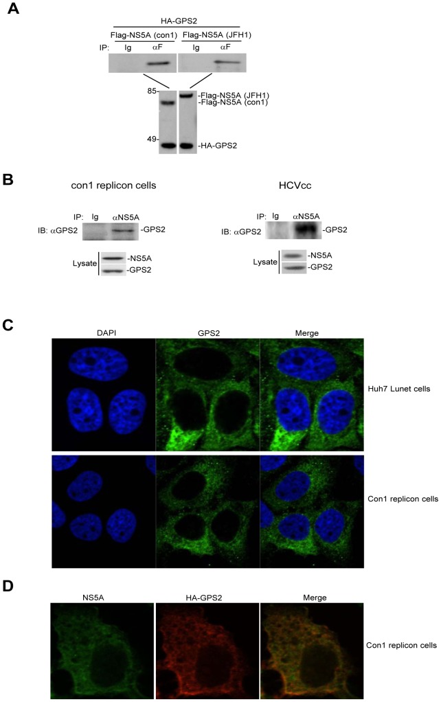
Figure 1. GPS2 interacts and colocalizes with NS5A.
(A) GPS2 interacts with NS5A in transfected cells. 293T cells (2×10⁶) were transfected with the indicated plasmids (6 µg each). Cell lysates were immunoprecipitated with anti-Flag (F) antibody or control mouse IgG (Ig). The immunoprecipitates were analyzed by Western blots with anti-HA antibody (upper panel). The expression levels of the transfected proteins were analyzed by Western blots with anti-HA or anti-Flag antibodies (lower panel). (B) GPS2 associates with NS5A under physiological conditions. Con1 replicon cells (1×10⁷) and Huh7.5.1 cells (2×10⁶) infected with JFH1 or left uninfected for 72 h were harvested. Cell lysates were immunoprecipitated with anti-NS5A antibody or control mouse IgG (Ig). The immunoprecipitates were analyzed by Western blots with anti-GPS2 antibody (upper panel). The expression of individual protein was analyzed by Western blots with the indicated antibodies (lower panel). (C) GPS2 mainly localizes in the cytoplasm in Huh7 Lunet cells and Con1 replicon cells. Huh7 Lunet cells and Con1 replicon cells were stained with antibody to GPS2. Alexa Fluor 488-conjugated anti-mouse secondary antibody were used to detect GPS2 (green). Cells were stained with DAPI to visualize the nuclei (blue). (D) GPS2 colocalizes with NS5A in Con1 cells. Con1 cells were transfected with HA-GPS2 and stained with antibodies to HA and NS5A 24 h later. Alexa Fluor 488-conjugated anti-mouse secondary antibody and Alexa Fluor 555-conjugated anti-rabbit secondary antibody were used to detect NS5A (green) and HA-GPS2 (red), respectively.

