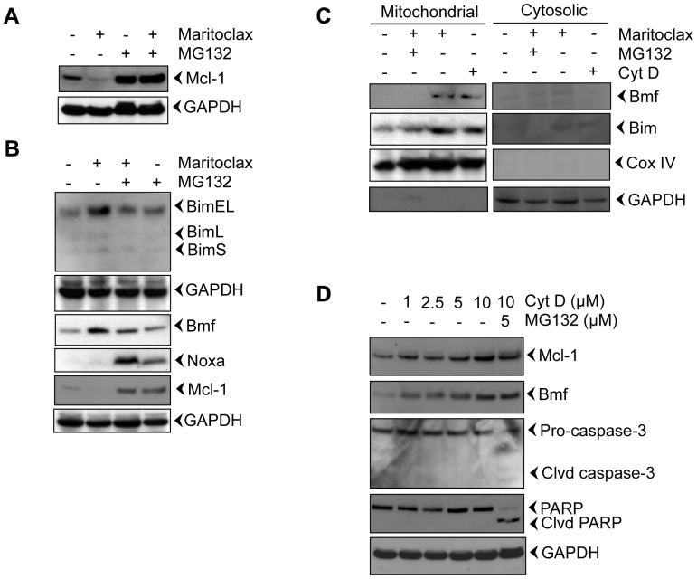
Figure 3. Maritoclax degrades Mcl-1 by proteasomal activation and releases Bim and Bmf from the cytoskeleton.
A-B, Melanoma cells were treated with 5.0 µM of Maritoclax alone or a combination of 5.0 µM Maritoclax and 5 µM MG132 for 12 h and levels of Mcl-1, Bmf, Bim, and Noxa were assessed by immunoblot analysis. C, Melanoma cells were treated with Maritoclax (5.0 µM, 12h) alone, a combination of Maritoclax (5.0 µM) and MG132 (5.0 µM) for 12 h, and cytochalasin D (10 µM, 3h) alone. Subcellular fractions were prepared. Levels of Bmf, Bim, Cox IV, and GAPDH were assessed by immunoblot analysis. D, Melanoma cells were treated with either indicated amount of cytochalasin D or MG132 for 12h. Whole cell extracts were prepared. Levels of Bmf, Mcl-1, Caspase-3, and PARP cleavage were assessed by immunoblot analysis. Loading was confirmed by GAPDH.

