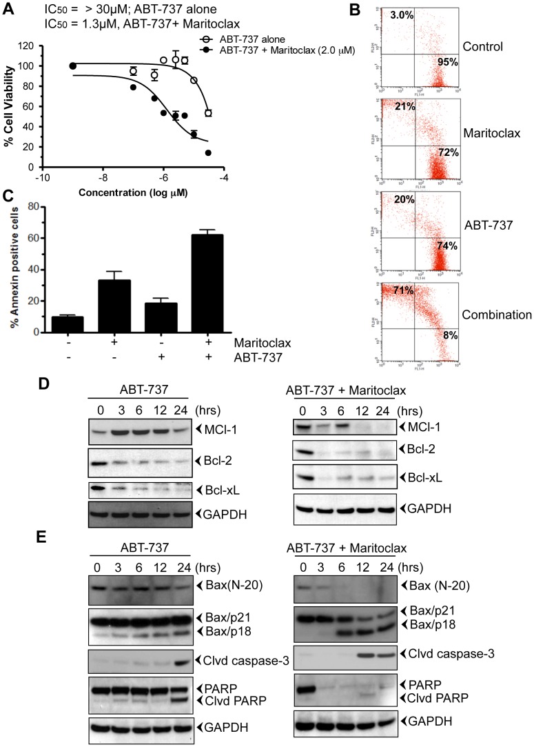
Figure 4. Maritoclax sensitizes melanoma cells to ABT-737.
A, UACC903 cells were treated with increasing amount of ABT-737 (0.1–30 µM) alone (open circle) or together with 2.0 µM Maritoclax (closed circle) and then incubated for 24 h. Cell viabilities were determined by MTT assay. B, UACC903 cells were treated with either 5.0 µM of ABT-737 alone or together with 2.5 µM Maritoclax and then incubated for 24h. Cells were stained with a Live/Dead assay reagent for 30 min and then analyzed through flow cytometer. C, Effect of Maritoclax on apoptosis. UACC903 cells were treated with either 5.0 µM of ABT-737 alone or in combination with 2.5 µM Maritoclax and incubated for 24h. Cells were stained with Annexin-V-FITC and then analyzed through flow cytometer. D-E, Melanoma cells UACC903 were treated with 5.0 µM of ABT-737 and 2.5 µM of Maritoclax together for mentioned time points. After incubation, cells were harvested; total lysates were prepared, and fractionated. The Western blot analysis was performed using mentioned antibodies and same blots were re-probed with either other antibodies or with anti-GAPDH to access loading.

