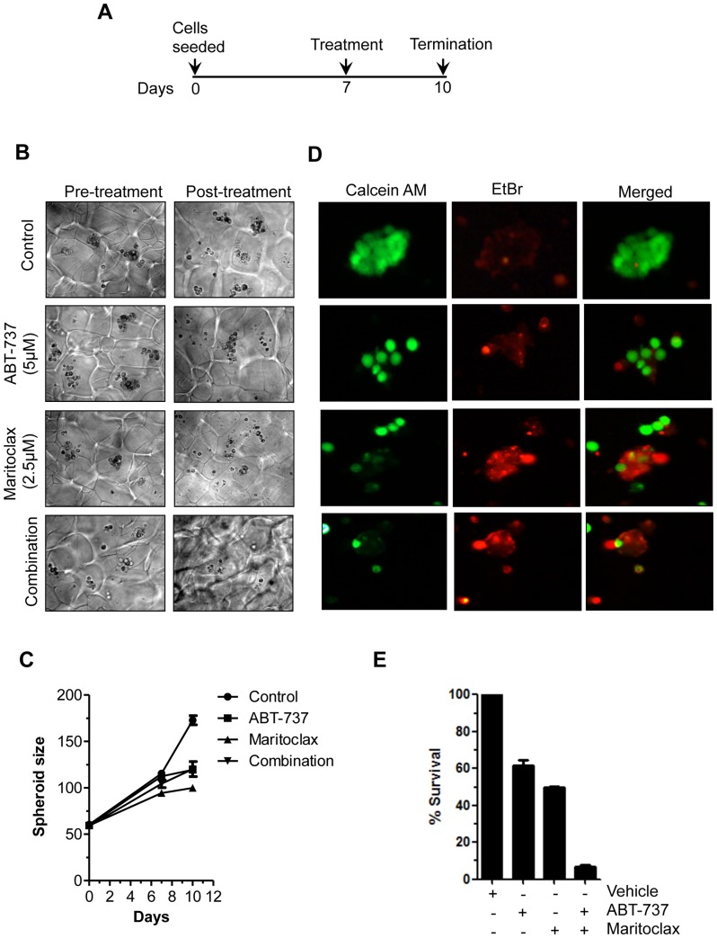
Figure 5. Maritoclax inhibits 3D spheroids and colony forming ability of melanoma cells.
A, UACC903 cells were grown as spheroids in Algimatrix for seven days and treated with either ABT-737 (5.0 µM) or Maritoclax (2.0 µM) alone or in combination for 3 days and on 10th day experiment was terminated. B-C, After termination of experiments spheroids were counted, size of spheroids was measured by using Image J software as described under Materials and Methods. Graph show corresponding quantification of spheroid growth using image analysis. D, Spheroids were isolated from matrix using dissolving buffer. Note the decrease of viable cells (calcein-AM) and increase of dead cells (ethidium bromide) in the treated spheroids only. E, UACC 903 cells were exposed to either 5.0 µM of ABT-737 or 2.5 µM Maritoclax alone or combination for 24h. Cells were re-plated after treatment for clonogenic assay. The effect of treatment was assessed on the basis of percent cell survival in comparison with the controls.

