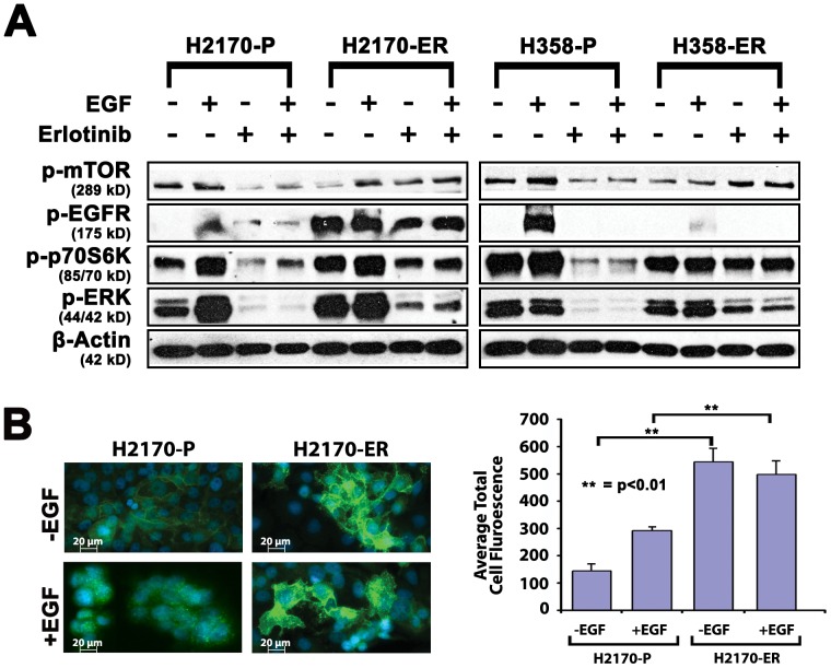
Figure 2. Differences in protein expression between parental and erlotinib resistant cell lines by western blotting.
A. EGFR is autophosphorylated in ER H2170 and downregulated in H358-E4 resistant cell lines. p-mTOR (S2448) and its downstream signaling protein phospho-p70S6K (T389) are upregulated in both resistant cell lines. H2170 and H358 parental and resistant cell lines were starved overnight in 0.5% BSA and then treated with or without 7.0 µM of erlotinib for 24 hours and cells were stimulated with 10 ng/mL of EGF for 2.5 minutes. Higher concentrations of erlotinib were used since these NSCLC cell lines have no EGFR TK mutation. Autophosphorylation of EGFR on Y1068 was seen in the absence of EGF in ER H2170 cells which was not seen in ER H358-E4 cells. Upregulation of p-mTOR and its downstream protein phosho-p70S6K (T389) is seen in H2170 resistant lines +/− erlotinib. ER H2170 cells show increased EGFR phosphorylation +/− EGF. Upregulation of p-ERK (2–5-fold) was also seen in ER H2170 and H358 cells in +/− erlotinib B. To confirm autophosphorylation of EGFR, cells were plated on chamber slides, allowed to adhere for 24 hours and then starved overnight. Cells were then treated with +/− EGF for 15 minutes, fixed with acetone: methanol and visualized with p-EGFR (Y1068) primary antibody and anti rabbit DyLight secondary antibody (Thermo Fisher Scientific) (green) or Hoechst dye for nuclear staining (blue) on a Zeiss Axio Observer Z1 fluorescent microscope. Graph showing relative average total cell fluorescence units per 8 microscopic fields. There was a 3.8-fold increase in fluorescence when comparing parental to resistant cells in the absence of EGF in H2170 cells.

