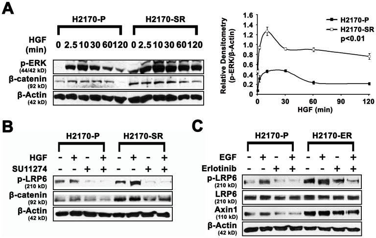
Figure 4. Differential expression of ERK/Wnt pathway proteins in parental and SU11274/Erlotinib resistant H2170 cells by western blotting.
A. In SR H2170 cells, HGF induced pronounced p-ERK signaling compared to parental cells. Cells were starved for 48 hours and then stimulated with 40 ng/mL of HGF. Western blotting in SR H2170 indicated that, HGF activated p-ERK (T202/Y204) remained high for 120 minutes compared to parental lines. Basal levels of active β-catenin were also 2-fold higher and remained high (3.6-fold) for 120 minutes after HGF treatment in SR H2170 cells compared to those in parental cells over 60 minutes incubation. These experiments were done in triplicate. Relative densitometry of p-ERK/β-actin in SR H2170 cells was depicted which is an average of three independent experiments (n = 3, p<0.01). B. Regulation of proteins in the Wnt signaling pathway after treatment of H2170 with SU11274. Upregulation of pLRP6 (2 to 3.0-fold) and β-catenin (3 to 8.0-fold) were seen in resistant H2170 cells in the presence or absence of SU11274. C. Regulation of proteins in the Wnt signaling pathway after treatment of ER H2170 cells with erlotinib. Upregulation of LRP6 (2 to 5-fold), and Axin1 (2 to 3.5-fold) were seen in resistant H2170 cells in the presence or absence of erlotinib.

