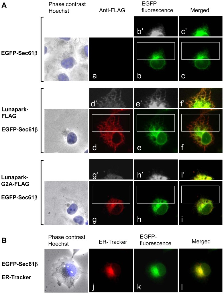
Figure 6. Protein Lunapark mainly localized to the peripheral ER and induced the ER morphological change in an N-myristoylation-dependent manner.
A. EGFP-Sec61β cDNA alone, Lunapark-FLAG cDNA and EGFP-Sec61β cDNA, or Lunapark-G2A-FLAG cDNA and EGFP-Sec61β cDNA, were transfected in to HEK-293T cells, and distribution of these proteins was evaluated by immunofluorescence analysis or fluorescence microscopic analysis. b′, c′, d′, e′, f′, g′, h′, and i′ show a close-up and over-exposed image of the area surrounded by a white box in b, c, d, e, f, g, h, and i, respectively. B. EGFP-Sec61β cDNA was transfected in to HEK-293T cells and the distribution of the protein was evaluated by fluorescence microscopic analysis. ER was detected with ER-Tracker Red.

