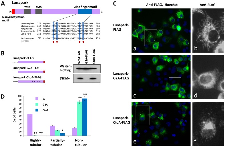
Figure 7. Role of the zinc finger motif of protein Lunapark in the ER morphological change induced by protein Lunapark.
A. Alignment of the zinc finger motif of Lunapark protein family members. Highly conserved cysteine residues are indicated by red arrows. B. Detection of protein N-myristoylation of Lunapark-CtoA-FLAG expressed in HEK293T cells. cDNAs encoding Lunapark-FLAG, Lunapark-G2A-FLAG, and Lunapark-CtoA-FLAG were transfected in to HEK293T cells, and their expression and the N-myristoylation of the products in the total cell lysates were evaluated by Western blotting analysis and [3H]myristic acid ([3H]Myr) labeling, respectively. C. Intracellular localization of Lunapark-FLAG, Lunapark-G2A-FLAG, and Lunapark-CtoA-FLAG was determined by immunofluorescence staining of HEK293T cells transfected with cDNAs encoding these three proteins using an anti-FLAG antibody. Right panel shows a close-up view of the area surrounded by a white box in the immunofluorescence image. D. Quantitative analysis of the ER morphological change in HEK293T cells induced by Lunapark-FLAG (WT), Lunapark-G2A-FLAG (G2A), and Lunapark-CtoA-FLAG (CtoA). cDNAs encoding Lunapark-FLAG, Lunapark-G2A-FLAG, and Lunapark-CtoA-FLAG were transfected in to HEK293T cells and the morphological change of the ER in each cell was determined by immunofluorescence staining and the extent of the ER morphological change was compared. The extent of ER morphological changes is expressed as a percentage of the number of cells with highly tubular, partially tubular, and non-tubular ER against the total number of transfected cells. Data are expressed as mean ± SD for five independent experiments. **P<0.001 vs. WT. *P<0.01 vs. WT.

