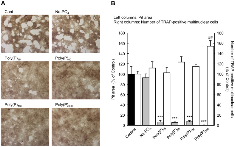
Figure 3. Inhibition of osteoclastic resorption activity by poly(P).
The osteoclast precursor cells were plated in calcium phosphate-coated plates (Bone Resorption Assay Plate 24) and stimulated with M-CSF and RANKL. After 3 days in culture, the cells were treated with the indicated lengths of poly(P) (1 mM) and incubated for an additional 2 days (for TRAP staining) or 4 days (for resorption activity assay). (A) Images of pits obtained by bright field microscopy. (B) The pit (white) area measured by image analysis. Total white areas visualized by bright microscopy were summed using image analysis software (Image J). The number of TRAP-positive multinuclear cells was counted after the cells were stained for TRAP activity. Values are expressed as means±SD. ***p<0.001, significantly different from the pit area of control. ## p<0.01, significantly different from the number of TRAP-positive cells of control (ANOVA with Bonferroni's post-test).

