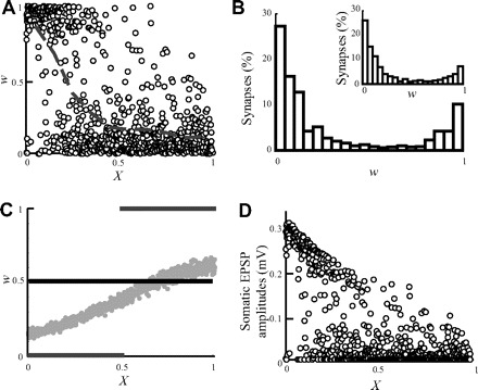
FIG. 1.

“The problem” with spike-timing–dependent plasticity (STDP) in dendrites. Following STDP, the strength of distal synapses diminishes, whereas that of proximal synapses is enhanced. A: w as a function of electrotonic distance from the soma at steady state following STDP. The gray line depicts the average w (computed at 0.1 λ bins). Strong synapses (w > 0.5) almost vanish for X >0.5. B: dendritic distribution of w values at steady state is bimodal. Inset: STDP in an isopotential compartment with the same membrane capacitance and conductance as in the dendritic model. C: 3 initial conditions for w were examined: w was set initially to 0.5 for all synapses (solid black line); w was arranged such that all synapses had the same efficacy at the soma (gray dots, see methods); w = 0 for synapses at 0 ≤ X < 0.5 and w = 1 for 0.5 ≤ X ≤1 (gray lines). The steady-state weight distribution is identical for these different initial conditions and is shown in A. D: somatic excitatory postsynaptic potential (EPSP) amplitudes as a function of distance from the soma in steady state.
