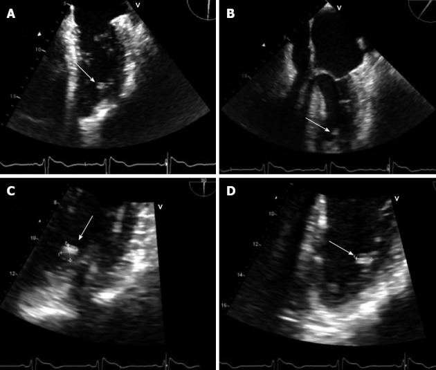
Figure 1.

Transesophageal echocardiogram. A: Mid esophagus view of the transesophageal echo (TEE) revealing left ventricular myxoma; B: TEE, four chamber view showing Left ventricular myxom; C: Magnified view showing a myxoma, size 1 cm x 2 cm; D: Magnified view showing a myxoma of 1 cm in horizontal direction. Showing its close proximity to trabecular muscles.
