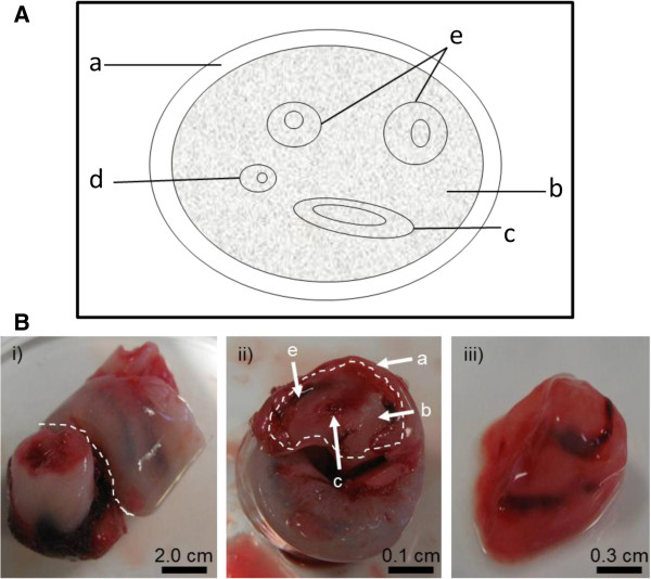
Figure 1.
Isolation of Umbilical cord tissue for DNA extraction. (A) A schematic structure of the umbilical cord (cross sectional view). a maternal sheath, b Wharton’s jelly; c umbilical vein; d allantoic duct; e umbilical arteries. (B) Umbilical cord preparation for gDNA extraction. i) Cord tissue was cut across as indicated with the white line. ii) Cross-section of the umbilical cord. The outer maternal sheath was removed as indicated with the white line. iii) The internal Wharton’s jelly with umbilical vein and arteries was used for gDNA extraction.

