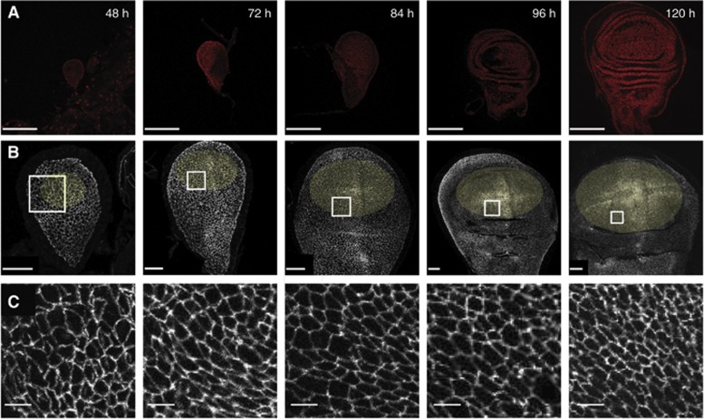
Figure 2.
Wing disc development. Confocal micrographs of wing discs fixed at the indicated ages after egg laying (AEL). (A) Hoechst staining labels nuclei. Scale=100 μm. (B) Wing discs expressing E-cadherin::GFP at endogeneous levels, marking the adherens junctions to show the apical cell geometries. Scale=20 μm. Yellow ellipses mark the areas of wing discs used for analysis. For 48–72 h wing discs, the Nubbin expression domain is used (Supplementary Figure S2), for older wing discs, an elliptical zone up to the first visible fold is used. (C) A magnified view of the white-square region marked in (B), scale=4 μm. Note that folds in the surface of the wing disc appear at ∼80 h AEL.

