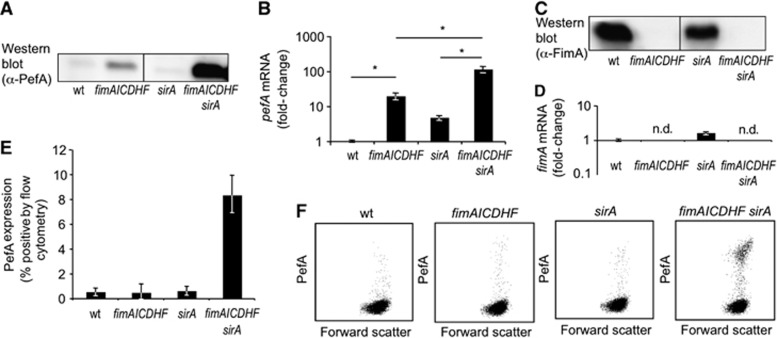
Figure 1.
SirA and the fimAICDHF operon synergize in repressing PefA expression. (A) Expression of PefA was detected in cell lysates of the indicated strains using western blot. (B) Relative expression of pefA-transcripts was assessed in RNA isolated from the indicated bacterial strains by real-time PCR. Bars represent the average of four independent experiments ±standard error. *P<0.05 (Student’s t-test). (C) Expression of FimA was detected in cell lysates of the indicated strains using western blot. (D) Relative expression of fimA-transcripts was assessed in RNA isolated from the indicated bacterial strains by real-time PCR. Bars represent the average of four independent experiments ±standard error. n.d., not detected. (E) Surface expression of PefA was detected by flow cytometry in the indicated bacterial strains. Bars represent the average of four independent experiments ±standard error. (F) Representative images of PefA expression detected by flow cytometry. wt, S. typhimurium wild type (SR-11); sirA, S. typhimurium sirA mutant (TS23); fimAICDHF, S. typhimurium ΔfimAICDHF mutant (SPN342); fimAICDHF sirA, S. typhimurium sirA ΔfimAICDHF mutant (TS24).
Source data for this figure is available on the online supplementary information page.

