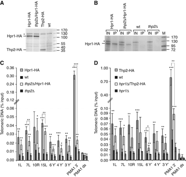
Figure 2.
Hpr1 is present at telomeres in thp2-Δ cells and Thp2 is present at telomeres in hpr1-Δ cells. (A) Deletion of THP2 does not influence the amount of Hpr1-HA in whole-cell protein extracts. Whole-cell protein extracts were prepared from strains grown to exponential phase in YPD at 30°C. Western blot analysis determined the amount of Hpr1-HA and Thp2-HA protein. An unspecific band is marked by an asterisk and was used as a loading control. Marker is given in kDa. (B) Deletion of THP2 does influence the specific pull-down efficiency of Hpr1-HA in ChIP experiments as determined by western blotting. After crosslink reversal, input (IN) and immunoprecipitates (IP) were analysed by PAGE. The Hpr1-HA protein band is highlighted by a line; an unspecific band is marked by an asterisk. Marker is given in kDa. (C) Hpr1-HA is associated with telomeres in thp2-Δ cells. Hpr1-HA and thp2-Δ/Hpr1-HA cells were grown to exponential phase, crosslinked and HA-associated chromatin was immunoprecipitated. The untagged wild-type (wt) and thp2-Δ strains served as negative controls. The immunoprecipitated telomeres 1L, 7L, 10R, 15L, 3*Y′, 4*Y′ and 6*Y′ were amplified by qPCR and are expressed as a percentage of the input. Amplification of the 3′end of PMA1 served as a positive control for Hpr1 association as in Figure 1. Hpr1 is not enriched at an untranscribed locus downstream of PMA1 (PMA1 ds, see Supplementary Figure S5B). Values of three independent biological replicates with standard deviation are shown. Statistical analyses are as in Figure 1. (D) Thp2-HA is associated with telomeres in hpr1-Δ cells. Thp2-HA and hpr1-Δ/Thp2-HA strains were grown and the HA-associated chromatin was detected as in (C). The wt and hpr1-Δ strains served as negative controls. Statistical analyses were calculated using the Student’s t-test (*P<0.05, **P<0.03 and ***P<0.01).

