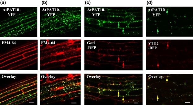
Fig. 10.

Subcellular localization of Arabidopsis AtPAT10 in the primary root. (a, b) Images were taken after 5 (a) and 60 min (b) of FM4-64 staining. After 60 min (b) AtPAT10-YFP (green) largely co-localizes with FM4-64 (red) in discrete punctae. (c) Some AtPAT10-YFP (green) co-localizes with the Golgi stack marker Got1 (red). Yellow arrows indicate co-localization between AtPAT10-YFP (green arrows) and Got1 (red arrows). (d) Some AtPAT10-YFP (green) co-localizes with the TGN/early endosome marker VTI12 (red). Yellow arrows indicates co-localization between AtPAT10-YFP (green arrows) and VTI12 (red arrows). Bars, 20 μm.
