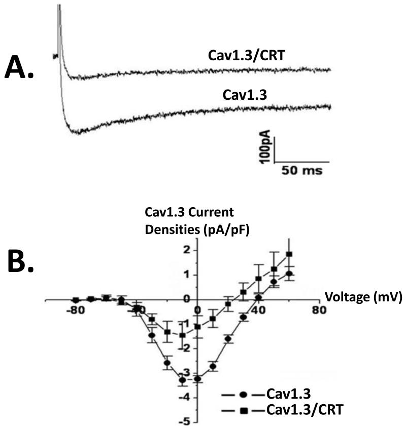
Figure4.
Functional Effect of co-expression of calreticulin with Cav1.3in tsA201 cells. Cav1.3 L type Ca current, ICa-L was recorded using whole cell mode of the patch clamp technique with 2 mmol/L Ca as a charge carrier. Panel A shows representative current tracings in the presence and absence of calreticulin. Note the decrease in basal current amplitude when calreticulin is present (Panel A). Panel B shows an current-voltage relationships of Cav1.3 ICa-L recorded by depolarizing pulses between −80 mV and +60 mV from a holding potential of −100 mV in cells transfected with Cav1.3 or Cav1.3/calreticulin. Functional Cav1.3 ICaL was recorded in the presence of the accessory subunits β2a and α2δ. CRT denotes calreticulin.

