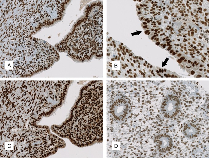
Figure 1.
Expression of HDAC in endometriosis and normal endometrium. Immunohistochemical expression patterns of HDAC-1 ((A) ×200; (B) ×400, with arrows marking the endometriotic epithelium) and HDAC-2 ((C), ×200) in ovarian endometriosis. HDAC-3 displayed a similar staining pattern as HDAC-2 and is therefore not represented. D, (×200) shows the expression of HDAC-1 in normal endometrium. HDAC indicates histone deacetylases.

