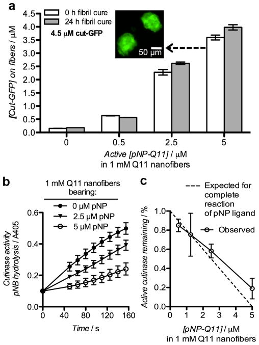
Figure 2. Covalent capture of cut-GFP by pNP-bearing Q11 nanofibers.
a) Conjugation of cut-GFP to nanofibers bearing different amounts of pNP-Q11, which were reacted with cut-GFP either immediately upon hydration or after extended assembly in water for 24 h. Immobilized cut-GFP was measured directly by GFP fluorescence on sedimented and resuspended nanofibers. Inset: Fluorescent micrograph of fibril aggregates of the formulation indicated. b) Reaction progress curves illustrating the residual activity of cutinase after overnight reaction with Q11 nanofibers bearing different amounts of pNP-Q11, as measured by p-nitrophenyl butyrate hydrolysis. (c) Initial velocities (v0) derived from (b) were used to calculate the % of active cutinase remaining after conjugation (5 μM cut-GFP, 0–5 μM pNP-Q11). All data show means ± s.d., n=3. pNP amounts are reported as the concentration of the enantiomerically active species (half of the total pNP racemate).

